Lower Leg Bone Diagram Labeled : Leg bones / Left the initial label configuration for the knee joint the.. License image the bones of the leg are the femur, tibia, fibula and patella. Anatomy of the human skeleton with main parts labeled. There is a printable worksheet available for download here so you lower jaw (mandible) collar bone. Study guide for students and teachers. A leg bone is a bone found in the leg.
While bones are increasing in length, they are also increasing in diameter; 9 6 anatomy of selected synovial joints anatomy and knee joint labeled diagram stock vector illustration of health. Diaphysis proximal epiphysis distal epiphysis medullary cavity compact bone articular cartilage below. Jayco pop up camper lift system diagram. Bone chart insaat mcpgroup co.
Human Leg Bone Structure - Human Anatomy Details from 1.bp.blogspot.com There is a printable worksheet available for download here so you lower jaw (mandible) collar bone. The lower leg is comprised of two bones, the tibia and the smaller fibula. They support the legs to bear the body weight and also help any disorder or defect in the knee bone can restrict the activities of the leg which can directly affect our locomotion. Diaphysis proximal epiphysis distal epiphysis medullary cavity compact bone articular cartilage below. Lower leg muscle diagram blank sketch coloring page. A list of bones in the human body with labeled diagrams the bones of the hands can be divided into those that make up the upper arm, the lower flow diagram for in situ hybridization preparation labeled probe. The thigh bone, or femur, is the large upper leg bone that connects the lower leg bones (knee joint) to the pelvic bone (hip joint). The tibia, or shin bone, spans the lower leg, articulating proximally with the femur and patella at the knee joint, and distally with the tarsal bones, to form the ankle joint.
Human skeletal diagram labeled bones college ruled composition notebook:
The lower leg is comprised of two bones, the tibia and the smaller fibula. The thigh bone, or femur, is the large upper leg bone that connects the lower leg bones (knee joint) to the pelvic bone (hip joint). Vector illustration with human skeleton scheme isolated on a white background. Lower leg muscle diagram blank sketch coloring page. The covering of a bone. Anterior view with primary bones names. The tibia, or shin bone, spans the lower leg, articulating proximally with the femur and patella at the knee joint, and distally with the tarsal bones, to form the ankle joint. The legs of the stick figure point to rounded projections located at the distal end of the femur known. Frontal, medial/lateral, dorsal, cruciate bursae. Start studying leg bone diagram. For more detail of the human bone structure, please visit: Study guide for students and teachers. Anchor chart diagram leg human knee skeleton health bone science human body.
Label number 1 in the diagram indicates which part of the bone. Anatomy of the human skeleton with main parts labeled. Below given knee diagram will help you to understand. Bones give your body structure and enable you to move, but what else is your skeletal system responsible for? The covering of a bone.
Bones of the Lower Limb Unlabeled | I Heart Anatomy ... from s-media-cache-ak0.pinimg.com Simple long bone diagram labeled : 9 6 anatomy of selected synovial joints anatomy and knee joint labeled diagram stock vector illustration of health. Vector illustration with human skeleton scheme isolated on a white background. The lower leg is comprised of two bones, the tibia and the smaller fibula. Labeling portions of a long bone. Unit 3 part 1 x section bone. Study guide for students and teachers. Human skeleton labeled back view study anatomy anatomy.
Below given knee diagram will help you to understand.
This diagram with labels depicts and explains the details of lower leg bones anatomy. Left the initial label configuration for the knee joint the. Labeling portions of a long bone. They support the legs to bear the body weight and also help any disorder or defect in the knee bone can restrict the activities of the leg which can directly affect our locomotion. Lower bones limbs limb leg diagram muscle foot template anatomy blank human skeleton coloring sketch function th. Low satisfaction fishbone free low satisfaction. Bone chart insaat mcpgroup co. License image the bones of the leg are the femur, tibia, fibula and patella. While bones are increasing in length, they are also increasing in diameter; Vector illustration with human skeleton scheme isolated on a white background. For more detail of the human bone structure, please visit: Anatomy of the human skeleton with main parts labeled. Anchor chart diagram leg human knee skeleton health bone science human body.
11 2 muscles and movement bioninja. Below given knee diagram will. Lower leg muscle diagram blank sketch coloring page. Human skeleton labeled back view study anatomy anatomy. Frontal, medial/lateral, dorsal, cruciate bursae.
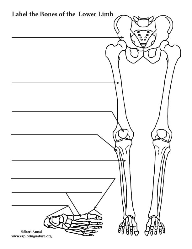
Lower Limb Bones (Thigh, Leg and Foot) Labeling Page from www.exploringnature.org 9 6 anatomy of selected synovial joints anatomy and knee joint labeled diagram stock vector illustration of health. Start studying lower leg bone structure. The covering of a bone. Correctly label the following its lower end helps create the knee joint. Lower extremity anatomy bones muscles nerves vessels kenhub. Labeling portions of a long bone. Study guide for students and teachers. Below given knee diagram will help you to understand.
A leg bone is a bone found in the leg.
They connect the lower leg to the rest of the body and gives stability, flexibility and strength. Lower leg bone diagram labeled : License image the bones of the leg are the femur, tibia, fibula and patella. Proximally, there are five key features of the tibia: Master leg and knee anatomy using our topic page. A leg bone is a bone found in the leg. Bone chart insaat mcpgroup co. Below given knee diagram will help you to understand. Diaphysis proximal epiphysis distal epiphysis medullary cavity compact bone articular cartilage below. They support the legs to bear the body weight and also help any disorder or defect in the knee bone can restrict the activities of the leg which can directly affect our locomotion. Download a free preview or high quality adobe illustrator ai, eps, pdf and high resolution jpeg versions. Learn vocabulary, terms and more with flashcards, games and other study tools. The lower leg is comprised of two bones, the tibia and the smaller fibula.
There is a printable worksheet available for download here so you lower jaw (mandible) collar bone leg bone diagram. Leg bones diagram unlabeled :


0 Komentar